India Pharma Outlook Team | Friday, 21 March 2025

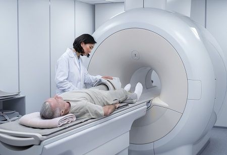
A new approach has made it possible for ultra-powerful 7T MRI scanners to reveal fine brain differences responsible for therapy-resistant epilepsy, presenting new prospects for patients. In a first-time study that has taken place at Addenbrooke's Hospital, Cambridge, the aforementioned methodological development has guided doctors to identify the areas of the brain that need treatment with surgical interventions for a potential cure.
7T MRI scanners work with a 7 Tesla magnetic field strength, which is more than twice the 3T scanners commonly used in clinical practice, and have improved imaging resolution. However, they have suffered from signal deficiencies within specific regions of interest within the brain, including the temporal lobes—the origins of epilepsy in many cases.
Researchers from universities in Cambridge and Paris have developed a strategy to solve the problem, and the findings went into Epilepsia. Focal epilepsy affects an estimated 360,000 people in the UK, many of whom are followed by neurology department - with a third continuing to have seizures, despite treatment with medications.
In such cases, surgery is the only potential cure. In order for surgery to be effective, it relies upon the identification of diseased brain tissue, and obtaining good visibility of the lesions on MRI can increase the chances of being seizure-free after surgery by 2 times.
Most NHS hospitals utilize 1.5T or 3T MRI scanners, which may be insufficient in detecting small but important lesions in drug-resistant epilepsy. Although 7T scanners show promise with more detail, their reliability is hindered by signal dropout.
This prompted researchers at the Wolfson Brain Imaging Centre in Cambridge and Université Paris-Saclay to try 'parallel transmit' technology. They were able to replace one transmitter with eight, which helped to eliminate blackspots in signal loss. This has improved the detection of lesions and could potentially improve outcomes for epilepsy surgery.